|
|
1.2 Diffraction contrast (BK)
|
Delicate brightness differences with spatial appearance (similar to interference contrast, DIK) down to dark field effect in relation to the degree of diaphragming. The applicability as normal bright field
objective is not influenced thereby. Acting as a convergence diaphragm, a wire diapragm that is lowered into the objective collects diffraction patterns that are generated by stopping down the light field or
condensered convergence diaphragm diminishes the optical resolution at least no more than e.g. the phase contrast method. Opposite to the stopped down bright field, interference even sharpens the image.
The method can be used for all objectives, the best results being from the objective 25x up.
| Objective |
Wire thickness |
 |
| 2,5 - 3,2x |
2mm |
| 10x |
1mm |
| 25x |
0,7mm |
| 40x |
0,5mm |
| 65 - 80x |
0,2mm |
| 100x |
0,1mm |
| generally: |
D=0.2/n.a. |
D=wire whickness
n.a.=numeric aperture |
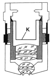
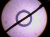
Convergence diaphragm inside the objective, central mounting 4-5 mm above the upper objective lens; fixing e.g. with 2-component glue.
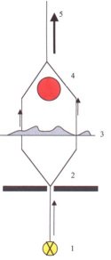 |
The light cone (1) is stopped down. Hereby, diffraction at the narrow slit (2) is induced. The secondary waves
generated hereby experience phase delay according to the specimen's density (thickness, refractive index???). Diffraction at the wire diaphragm results in lighting or darkening in correspondence to the run
length difference at the reunion of the secondary waves. The microscopic image shows a contrast enhancement in proportion to the degree of stopping down. |
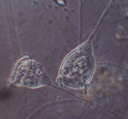
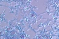
Vorticella campanula, objective 40x |
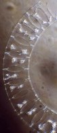
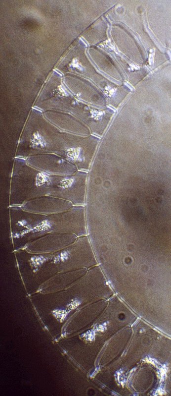
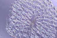
Eucampia (diatom, North Sea), objective 40x |
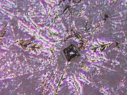
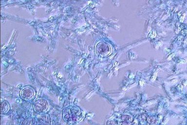
Crystal growth of table salt, objective 10x |
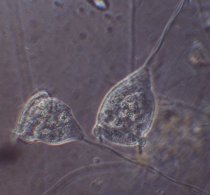
Experiment: Fascinating zooplancton
Material: objective (e.g. 40x), wooden toothpick, ruler, copper wire of thickness 0.3mm, tweezers, small pliers, 2-component glue, zooplancton (paramecia, vorticella, etc.).
Realization:
Sense the depth from the objective's upper rim to the lens by careful use of the toothpick and measure with the ruler. Subtract 5mm. Next, measure the objective's upper diameter. Bend a wire bow
according to the image, lower it into the objective and glue it to the upper objective rim. After the glue has cured, center the wire bow using the toothpick. Viewing down the eyepiece tube after
removing the eyepiece and screwing in the objective, the centering of the wire diaphragm can be controlled and adjusted if necessary.
Exercises:
a) Microscopize using the modified objective with open
b) partially and completely stopped down condenser diaphragm
c) Repeat with lowered condensor.
Further informations about diffraction contrast: Mikrokosmos (german), Edition 1/1999 and Edition 5/1999
Comparing images shot at the University Reutlingen's Institute for Applied Research using a Zeiss Axioplan microscope showed hardly noticeable
differences between differential interfence contrast and diffraction contrast, which justifies the term 'highlight' for this invention. The optical industry so far ignores the invention, not taking into account
that by modification of existing objectives the big pools of schools and high schools as well as hobby microscopists could be accessed. Specially built diffraction contrast objectives could be marketed, too.
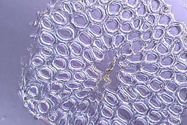
Cross section, Neofluar objective 40x difference interference contrast, DIK |

Cross section, Neofluar objective 40x, BK |
Cross section coco fiber, Neofluar objective 40x difference interference contrast, DIK |
Cross section coco fiber, Neofluar objective 40x, BK |
 |
Phase contrast objectives offer an interesting possibility: by additionally inserting a convergence diaphragm,
it is possible not only to microscopize in phase contrast or dark field, but also in diffraction contrast. This is achieved by simply switching phase contrast to brieght field and stopping down accordingly.
|
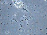
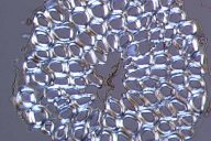
Lactic acid bacteriae in yoghurt, objective PH40x |
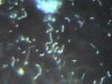
Same objective, in dark field: blurring from irradiation |
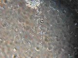
Same objective, in diffraction contrast |
 Top Top
 Home Home
|